ExtraCORPOREAL membrane oxygenation (ecmo)
What is it?
Types of ECMO
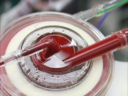
It is an extracorporeal perfusion technique that provides oxygenated blood to the patient. Can be a short or long term solution. The ECMO circuit is composed by: a centrifugal pump, an oxygenator, cannulas and a heater cooler.
Prior on initiating the ECMO, cannulas are placed in the patient vein or arteries and are connected to the circuit. Once the medical team are ready, the perfusionist activates the ECMO machine. The patient’s blood is drained from the venous circulation. The blood reaches the centrifugal pump and it is pushed into the oxygenator. Here the blood gets oxygenated, warmed up and retuned back into the patient blood circulation.
The perfusion manages the amount of oxygen and air flow delivered to the oxygenator to reach an appropriate exchange of O2 and CO2.
Types of ECMO
Types of ECMO

- Veno-Arterial (VA) ECMO:
the VA ECMO drains venous blood, oxygenates the blood and pumps it back into an artery. This type of ECMO provides support to the heart and lungs.
Cannulation sites:
- Femoral Vein (drainage) and Femoral Artery (return)
- Central VA ECMO, Right Atrium (drainage) and Aorta (return)
- Veno-Veno (VV) ECMO:
the VV ECMO drains desaturated venous blood from the patient, oxygenates the blood and pumps the blood back into a venous compartment. This type of ECMO provides support to the lungs only.
Cannulation sites:
- Femoral vein (drainage) and Jugular vein (return)
- Femoral - Femoral vein
- Jugular vein (double lumen cannula)
When is it used?
When is it used?

VV ECMO
- Pneumonia
- ARDS (Acute respiratory distress syndrome)
- Pulmonary contusion
- Status asthmaticus
- Aspiration or inhalation injury,
- Drowning
VA ECMO
- Cardiac arrest or cardiogenic shock
- Poisoning and drug overdose
- Pulmonary embolism
- Hyperthermia
- Massive pulmonary haemorrhage
- Bridge to: transplant, recovery, decision
- Weaning from bypass
- Sepsis
ECMO played an important role in treating patients affected by COVID-19 virus.
Ventricular assisted device (VAD)
What is it?
Types of VAD

Ventricular Assisted Device (VAD) is an extracorporeal technique that is used to provide mechanical support to the patient heart.
There are different types of VAD:
Left, Right and bilateral VAD (BIVAD). This VADs can be temporary or permanent.
- Left Ventricle VAD: helps the left ventricle to pump blood back into the aorta.
- Right Ventricle VAD: helps the right ventricle to pump blood into the pulmonary artery. Sometimes an ECMO can be used as RVAD.
- BIVAD: gives mechanical support to both right and left ventricles.
Types of VAD
Types of VAD

Transcutaneous VAD
In this type of VAD the pump and the device controller is located outside the patient body. The pump is connected to the patient via cannulas. For the LVAD the cannulas are located in the left ventricle (drainage) and the aorta (return). For the RVAD the cannulas are located in the right ventricle (drainage) and the pulmonary artery (return).
Implanted VAD
In this VAD the pump is located inside the patient and the device controller out side the patient. The pump is connected into the left ventricle and a driveline will link the pump to the device controller.
When is it used?
When is it used?
- Bridge to transplantation
- Bridge to recovery
- End-stage heart failure
- Bridge to destination therapy
- Weaning from cardiopulmonary bypass